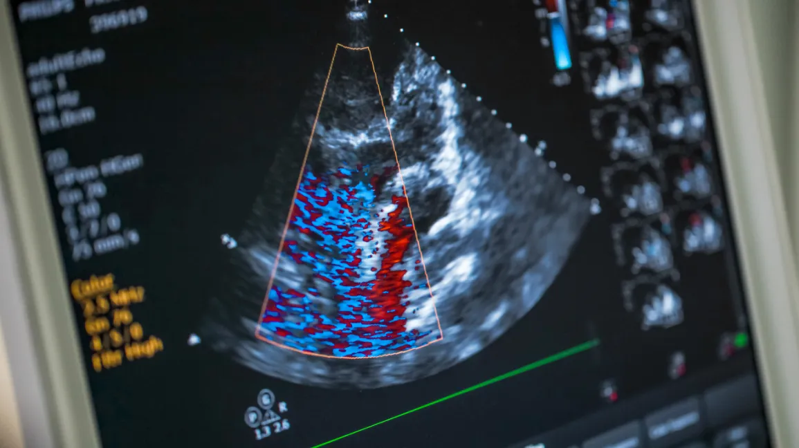
2-Dimensional Echocardiogram (Cardiac Ultrasound)
This is a safe and painless procedure that uses a transducer (a small, microphone-type device) to send high-frequency sound waves to the heart. The technologist can view the heart valves and pumping strength, look for structural defects, heart enlargement and/or fluid buildup. A Doppler exam and Color Flow exam are also performed. This allows doctors to evaluate blood flow through the heart. The technologist will videotape parts of the study for later diagnosis by the cardiologist.
4D Echocardiography - with new technology to new borders
The only current method in cardiology by which the heart can be studied with high spatial and temporal resolution in three dimensions in real-time.
Unlike sectional imaging technologies (CT and MRI) conventional echocardiography was unable to display neither spatial relationship to other structures nor multi-dimensional image representation. With the introduction of real-time 3D echocardiography, dimensional images of the heart, and especially of the heart valves, are now possible.
An ECG may be recommended if you are experiencing arrhythmia, chest pain, or palpitations and an abnormal ECG result can be a signal of several different heart conditions.